By Landau Buissereth, Biosafety Specialist
Histology specimen preparation in BSL-1 or BSL-2 labs using microtomes or cryostats involves very sharp blades around your fingers. Additionally, you can be exposed to infectious materials when working with unfixed biohazardous samples, which pose a much higher risk than fixed tissue.
Be Prepared
Read the operator manual for the microtome or cryostat and request training from your principal investigator, lab manager, or a colleague who has experience using the equipment. In addition, note whether your sample is fixed or unfixed and which biohazardous agent(s) it may harbor.
Properly Load and Unload Samples
Ensure samples are properly trimmed prior to mounting onto the stage. Cover the blade by using the provided finger guards or a protective foam guard over the blade during mounting/removal of a sample. Use forceps to remove the blade, and wear either cut-resistant gloves or two layers of nitrile gloves to help protect your skin. Recommended cut-resistant gloves include the following:
- Milwaukee Large Red Nitrile Level 1 Cut Resistant Dipped Work Gloves: https://www.homedepot.com/p/Milwaukee-Large-Red-Nitrile-Level-1-Cut-Resistant-Dipped-Work-Gloves-48-22-8902/303635833
- Ansell HyFlex™ 11-318 Lightweight Uncoated Cut-Resistant Gloves: https://www.fishersci.com/shop/products/ansell-hyflex-ultra-light-level-2-cut-resistant-nylon-gloves/19152304#?keyword=11-318
- VWR® PureTouch Cut-Resistant Glove Liners: https://us.vwr.com/store/product/22478406/vwr-puretouch-cut-resistant-glove-liners
Perform Routine Sanitation
Always clean the equipment between users and at the end of a session. A tuberculocidal disinfectant is recommended. Cryostats are more difficult to disinfect between samples since the cold can make the disinfectant build up on the knife, so it is best to finish the entire session and then perform a comprehensive decontamination by removing the stage and allowing it to come to room temperature before cleaning. Allow it to dry completely before returning it to the cryostat.
Cleaning the Microtome or Cryostat
Put on protective equipment including gloves (cut-resistant or double nitrile) and a lab coat. Use a pair of forceps to carefully remove the blade Either discard the blade directly in a sharps container or if re-using it, place in a container of disinfectant to soak. Ensure complete contact time, then use forceps and a handled brush to remove residue and scrub clean, followed by water rinse. Use forceps to remove residue, stray shavings, etc., from the interior of the microtome and place in a biohazard bag. Soak the interior components with disinfectant, applied with a squirt bottle. Use a cloth, manipulate with forceps where possible, to scrub and decontaminate. If needed, use a handled brush to access hard to reach areas and wear facial protection because a brush is more likely to cause splatter. Soak up residual disinfectant and rinse/dry with 95% ethanol. Let air dry.
Note on Bleach
The first impulse may be to use bleach (10% bleach followed by multiple rinses of 70% ethanol), but you can protect the blade by evaluating the agents most likely to be in the samples and choosing a less corrosive alternative disinfectant.
Things to Avoid
Do not leave the blade exposed during sample loading and unloading. This is one of the easiest ways to sustain an injury. Remember to cover the blade.
NEVER handle the blade with unprotected hands. Make sure to use forceps and wear gloves.
Always utilize Personal Protective Equipment (PPE) - gloves can minimize or possibly prevent cuts and lacerations.
Important Safety Considerations
Whenever possible, the use of fixed tissue will reduce potential exposure to pathogens.
Unfixed tissue potentially exposes the operator to pathogens due to the nature of the operation of the unit. The use of additional PPE (e.g., N95 respirator, eye and face protection), facility barriers (e.g., placement of equipment in a dedicated room with door that can be closed while work is progress), and administrative controls (e.g. restrict personnel) may be needed. Contact EHS to perform a risk assessment and coordinate respirator fit testing and training.
Technical Guidance
For technical guidance on microtomes, please see the following helpful resource from Leica Biosystems (Introduction to Microtomy: Preparing & Sectioning Paraffin Embedded Tissue): https://www.leicabiosystems.com/us/knowledge-pathway/steps-to-better-microtomy-flotation-section-drying/
Adapted from article originally written by Cornell University EHS

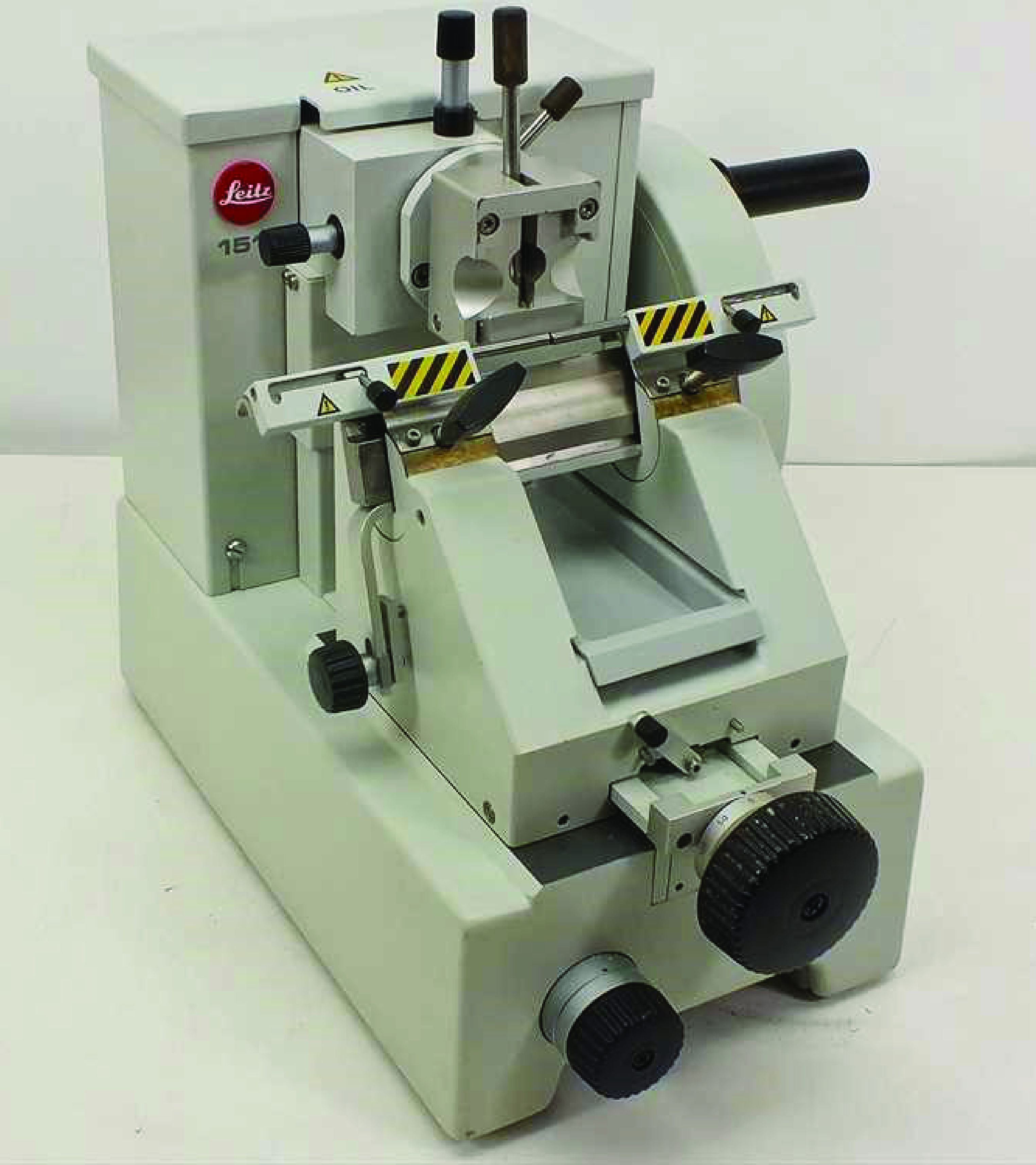
The microtome finger guards are engaged in this picture (marked with yellow and black stripes). Always make sure that you protect yourself from the knife blade by using the guards, a foam cover, or tongs, depending on the manipulations being performed.

The cryostat finger guard is located on the stage where the blade is mounted. When NOT sectioning, the blade cover should be flipped closed to protect from possible injury.

